Reef Alchemy by Randy Holmes-Farley

Sea Urchins: A Chemical Perspective


Most of my reef chemistry articles deal with issues with clear practical importance to aquarists, and that tendency will certainly continue in the future. These articles have included such topics as how to solve calcium and alkalinity problems and making and using additives. This article, however, is the first in a series that is intended to provide a chemical perspective on events that occur in reef aquaria. They may not cause aquarists to change their aquarium husbandry techniques, but they may allow a greater appreciation for the chemistry lying just under the surface of a reef aquarium.

This first article will examine sea urchins. Sea urchins are beautiful organisms, and many aquarists keep a variety of species in reef aquaria. Many fascinating articles have been written on them, often describing their physical attributes, their biological mysteries, and in some cases, techniques for aquarium husbandry. This article by Shimek, for example, is well suited as an introduction to these organisms.

Sea urchins, however, have a chemical side that may be of interest to many aquarists. Why are the spines colored? What are they made of? I always wondered what they were, but never knew until I wrote this article. Do sea urchins pull calcium and alkalinity out of the water, like corals? These are the sorts of questions that will be addressed in this article.

Sea Urchin Spines

The first thing that comes to mind when most aquarists hear the name sea urchin is an animal covered in spines. Sea urchin spines come in a wide range of sizes, shapes, and colors. Some urchin spines are long and slender (Figure 1). Others are short and fat (Figure 2). In still others, the spines have been almost eliminated (Figure 3). Many researchers in the past century have studied sea urchin spines, so quite a lot is known about them. They provide a fascinating look at how nature solves bioengineering problems, and they are a good place to start this chemical perspective on sea urchins.

Figure 1. An unidentified sea urchin, probably Arbacia punctulata, photographed
off the coast of Fort Pierce, Florida by Andrew K. Osborn.

Not all urchin spines are the same. They have, in fact, evolved in many different ways, as is clear in the photographs in this article. Some are covered in epidermis (tissue), while others, notably the pencil urchins (and all cidarid urchins), are not. Consequently, the discussions that follow are intended to highlight the chemical aspects of certain urchins, but the attributes detailed may not extend to any particular species.

Sea Urchin Spines: Structure

While sea urchin spines have a wide range of appearances, they also have some commonality. All, for example, are largely made of calcium carbonate, as are coral skeletons. Interestingly, recent studies have shown some sea urchin spines to be large single crystals of calcite,1 while others are mosaics of many individual crystals.2 For example, by using X-ray reflection, the spines of Heterocentrotus trigonarius have been shown to consist of 10-cm long single crystals of magnesium-rich calcite.1 Calcite is a particular crystalline form of calcium carbonate in which the calcium and carbonate ions are arranged in a slightly different pattern than in the other predominant form of calcium carbonate found in marine organisms: aragonite. Corals use aragonite for their skeletons, but urchins have found calcite to be more suitable for their spines. The magnesium-rich part of the description implies that a significant portion of the calcium ions have been swapped for magnesium ions, and the exact amount of magnesium present is discussed later in this article.

Figure 2. A sea urchin believed to be Eucidaris tribuloides, photographed
by Eric Edmondson (Spider_Whistle) in his aquarium.

Figure 3. A sea urchin believed to be Colobocentrotus atratus, photographed
by Bob Bottini (aquababy) owner of Tanks alot!

In all Heterocentrotus trigonarius spines, the calcite crystal is aligned in a particular direction in relation to the spine's axis. In other words, the arrangement of calcium and carbonate ions was perfectly aligned with respect to the length of the spine, leaving no gaps along which the spine might easily crack.2

The spine itself, however, is not a solid crystal, like a chunk of calcite that might reside in a rock collection. Rather, it is very porous, as seen by scanning and transmission electron microscopy (SEM, and TEM, respectively). These pores consist of two types: a continuous assortment of macropores that are easily seen by SEM, and a series of many small (80 nm) protein inclusions that are easily seen by TEM. These proteins are, in fact, critical for the spine's initial formation, and will be discussed later in this article.

Sea Urchin Spines: Composition

As mentioned above, the crystals are magnesium-rich calcite, containing 2-25 mole percent magnesium ions (75-98 mole percent calcium)2,3 This level of magnesium is appreciably higher than in most coral skeletons (which use the aragonite crystal form of calcium carbonate), and similar to or higher than many calcareous algae (which often deposit calcite having several mole percent magnesium). The magnesium content of the spines has been shown to vary somewhat with water temperature, and has also been shown to increase by about 2 mole percent from the tip of the spine to the base.

It has been suggested that the presence of magnesium in the calcite strengthens the calcite by altering the way cracks can propagate through it. The additional magnesium near the base makes the base stronger, thus increasing the likelihood that any spine that does break will break farther from the body, causing the urchin to lose less material than if the break were nearer to the body.2

When exposed to excessive heavy metals, the spines of sea urchins can incorporate these metals into their crystal structure. Lead and zinc, for example, have been shown to be incorporated into the spines of Paracentrotus lividus and Arbacia lixula when the ambient levels rise above those in normal seawater.4 What effect these incorporations have on the spines, for example, on their strength is not clear.

Sea Urchin Spines: Formation

A multitude of studies have examined the deposition of calcium carbonate in sea urchin larvae, and a few have studied it in adults. As might be expected from a process that results in a large, shaped single crystal, the deposition is very carefully controlled. How this happens is only poorly understood. As with calcification in corals, it is a very complicated process. It involves numerous proteins and other organic compounds. What is known of the overall process is outlined in the following sections.

Sea Urchin Spine Formation: Uptake of Calcium and Carbonate

Sea urchins can take calcium and alkalinity directly from the water column to deposit calcite to form spines. This has been demonstrated by the fact that some (e.g., Strongylocentrotus purpuratus) have been shown to be able to take calcium from solution and calcify even when kept under starvation conditions, although the calcification rate was slower than under fed conditions.5

At least two species, however, have been shown to have some propensity to consume solid calcium carbonate that may be dissolved by the lower pH in their guts and released to the organism to provide the building blocks for calcification, sort of like miniature CaCO3 reactors.6 In these experiments, Diadema setosum and Echinometra sp. were shown to prefer artificial foods with powdered calcite added over the same foods with no calcite. Many urchins might similarly obtain calcium and carbonate ("alkalinity") from the calcareous algae and other bits of calcium carbonate that they consume. [As an interesting aside, it has been claimed that many calcareous algae deposit CaCO3 in an effort to make themselves less palatable to fish.7 In the case of sea urchins, this strategy appears to backfire.]

Will urchins significantly deplete calcium and alkalinity in a reef aquarium? Probably not very much relative to other calcifying organisms, but in a new aquarium in which coralline algae and corals may be calcifying slowly, this relatively low depletion rate may be an appreciable part of the total. When I first set up my reef aquarium, I used wild Florida live rock that contained quite a load of sea urchin hitchhikers. More than 30 rock-boring urchins (Echinometra lucunter) were present, and they grew very rapidly for a year or so, before dwindling. During that time, coralline algae grew poorly (even in areas not browsed by the urchins) and I had few rapidly calcifying corals. Consequently, while I did not realize it at the time, some of the limewater that I was adding may have been ending up in the urchins. As an aside, studies have shown that moderate grazing by sea urchins can increase the spread of coralline algae from the chips that are released. However, in a finite space like a reef aquarium, heavy sea urchin grazing may overwhelm the growth of coralline algae.

Sea Urchin Spine Formation: The Role of Organics

The spicules in sea urchin embryos, as well as the spines in growing adults, are deposited by primary mesenchyme cells, which surround the growing tips of the spines. These cells accumulate calcium and secrete calcium carbonate.8 Exactly how the secreted calcium and carbonate ions become shaped into spines, however, is not entirely clear.

More than a dozen different proteins have been found to be associated with calcification in sea urchins.8-16 In the completed spine, many of the proteins remain trapped inside pores in the spine. In one case, the trapped protein was estimated to comprise 0.1% of the spine's total mass.3,8 In a related experiment, some of the proteins (acidic glycoproteins) present in sea urchin calcite were extracted and added back to calcite crystals in vitro (in test tubes) to see what effect it had. These proteins turned out to adsorb onto specific crystalline facets of the calcite where they altered crystallization along those facets.9 In another experiment, proteins extracted from the spines of Paracentrotus lividus also adsorbed onto only a single crystal facet, encouraging one of the other facets to grow more. Interestingly, when magnesium was added to the mix, the proteins directed growth to occur on a different facet.10 In this way, the sea urchin can use a variety of organic and inorganic materials to modify growth of spines to specific sizes and shapes as it "chooses," based on the materials secreted onto the growing surfaces.10,15

Sea Urchin Spines: Coloration

The descriptions of sea urchin spines provided to this point do not explain why they are colored. Indeed, the coloration of sea urchin spines has been the subject of numerous research projects for decades. Obviously, with so many different urchins of different colors, the explanations of the colors will vary, but sea urchins as a group have a relatively novel set of chromophores. In fact, many of these form a homologous family of chemical structures that has been named the spinochromes ('spino' for spine and 'chrome' for color). Figures 4 and 5 show some of these novel structures that have been isolated from sea urchins, but many related structures have also been isolated from them.

Each species of urchin may have many different chromophores, giving it a distinct color, just as mixing different pigments in paints gives different colors. The red sea urchin Strongylocentrotus franciscanus contains spinochrome B, echinochrome A and spinochrome E.17 Diadema setosum (which is dark purple/black) contains echinochrome A, spinochrome A and the methyl ether of echinochrome A.18 Strongylocentrotus droebachiensis (which can have red, green, or violet spines that are often white tipped: Figure 7) contains spinochromes A, C, D, and E as well as the two dimers shown in Figure 5.19 The spines of Strongylocentrotus nudus have been found to contain at least 11 different pigments, including spinochromes A, B, C, and E and echinochrome A.20

Figure 4. Some of the typical chromophores isolated from sea urchins.

By varying the substituents on these spinochrome rings, the urchins have made a variety of different colors, with only small chemical changes potentially leading to large color changes. For example, having hydroxyl groups on only the quinone side of the ring system (the side with the two C=O groups) often gives oranges and reds, while having hydroxyl groups on both rings gives purples and blues.21 Some of the –OH groups attached to the rings are also acidic, and can deprotonate to –O- groups. Echinochrome A, for example, has three of the –OH groups deprotonated under the pH and calcium concentrations in normal seawater (Figure 6).22 This deprotonation also drastically changes the color, and is especially likely in the presence of high calcium concentrations.

That said, why would a sea urchin care about color? After all, they don't "see" each other. Are these dyes even produced for the color effect, or for other reasons? Some may use color to warn potential predators if they are especially poisonous (discussed later in the article). Other urchins may use these molecules for purposes entirely different from color. In fact, the spinochromes may have evolved to perform several different functions for the urchins.

It has been noted, for example, that spines of many species of sea urchins are rarely covered with any epiphytic growth (that is, few or no organisms settle onto the spines to grow, although some do get such growth, as shown in Figure 8). It has been suggested that the spinochromes my provide part of this natural anti-biofouling coating. In fact, the spinochromes and related compounds have been shown to be effective anti-biofouling agents for fishing nets,23 and other applications.24-26 How can this inhibition work? The spinochromes are very water soluble molecules, and they could easily leak (or be secreted) from the various tissues containing them into the intercellular water of the epidermis as well as into the nearby water column. At adequate concentrations, these molecules could then kill organisms that try to settle onto this "toxic" substrate. At sublethal concentrations, the secreted spinochromes may simply irritate organisms sufficient to get them to move away (or not settle in the first place). This process has not, however, been proven to take place.

Figure 5. Chromophores isolated from Strongylocentrotus droebachiensis that are
dimers of spinochromes.

An alternate explanation of the advantages of the spinochromes involves protection from oxidation by superoxide free radicals (O2-). Such chemistry is well beyond the scope of this article, but these highly oxidizing free radicals have been implicated in all sorts of undesirable phenomena in organisms, including diseases in humans ranging from aging and cancer to heart disease. It also plays a role in certain types of coral bleaching. Such radicals are very reactive, and damage nearby organic molecules. The benefit to humans of the antioxidant vitamins, for example, may be that they rapidly scavenge free radicals.

In the case of sea urchins, they may use the spinochromes and related compounds to defend against the superoxide free radicals (O2-) that bombard them in the ocean.27-31 These oxidizing species are generated by physical processes involving oxygen and light, and also are made and secreted by many organisms. Several of the spinochromes have been shown to be powerful superoxide and reactive iron scavengers.32,33 Whether this is indeed an advantage that benefits sea urchins, or whether it is an unintended consequence of the chemistry used to attain some other goal, is not clear.

Figure 6. Echinochrome A shown in the protonated state (top) and the
deprotonated radical anion state complexed to calcium (bottom).

Finally, sea urchin embryos are easily damaged by ambient ultraviolet light.34 Consequently, it is possible that these pigments provide protection for the growing and adult urchins exposed to intense sunlight.

Figure 7. Strongylocentrotus droebachiensis. Photo by Ronald Shimek.

It is also possible that we do not yet know the most important function of the spinochromes, and that all of the effects described above are incidental to some unknown primary use.

Sea Urchin Teeth

Like sea urchin spines, sea urchin teeth have also received significant study.35-41 Sea urchins have five continuously growing teeth (Figure 10) with a complex mineral structure. Their teeth are amazingly rugged, and sea urchins have been known to eat through ferroconcrete piers, as well as lead ensheathed deep-water telephone cables. It turns out that their teeth are formed, like the spines, of high magnesian calcite (4.5 - 13 mole percent magnesium).35 For some urchins, the magnesium content varies considerably by location within the tooth, with the most magnesium concentrated at the tip, just the opposite of the magnesium distribution in spines.37,38 The tip of an urchin tooth undergoes extensive erosion, both mechanical and chemical. The mechanical erosion from constant chewing on tough foods may explain why an urchin would want the tip of the tooth to be as strong as possible, driving the development of higher magnesium calcite in tooth tips.

Figure 8. Zoanthids growing on the spines of the deep-water cidarid
urchin Stylocidaris lineata. Photo by Ronald Shimek.

The chemical aspects of tooth wear may also be important for some species. Strongylocentrotus droebachiensis, for example, eats certain kelp species in the genus Desmarestia that pack sulfuric acid into vacuoles (small reservoirs) in some of their cells. After grazing on such algae for a while the urchin teeth begin to dissolve and develop a gap between them. This gap allows the algal blades to more frequently slip out of their grip. I have not seen any analysis of the magnesium content of the teeth of these urchins, but high magnesium calcites are generally more soluble than lower magnesium calcites (or pure calcite), so one might predict that these urchins may have less magnesium in their teeth than other species.

In addition to the magnesian calcite, some parts of the tooth are amorphous plates and rods of calcium carbonate, although crystalline calcite predominates. In many cases, the calcite is in the form of long crystalline fibers that are thin at the base (1 micron) and thicker at the end (20 microns).

These calcite fibers have a thin organic coating (probably the proteins that control deposition and growth) that is, in turn, surrounded by polycrystalline calcite containing up to 35 mole percent magnesium. This structure has been referred to as a "gradient fiber-reinforced ceramic matrix composite, whose microhardness and toughness decrease gradually" from the tip to the base.38 Additionally, there are plates of calcite with an interior of amorphous calcium carbonate in the tooth that serve to prevent the propagation of tooth cracks.38

Overall, the teeth of sea urchins have a complex array of techniques for making them as hard and durable as possible, and they have served to inspire scientists and engineers trying to make similarly durable structures.33

Sea Urchin Venom

Many sea urchin spines (and also their pedicellariae, short pincers that they use for defense) contain venom that makes getting spined more painful than it might otherwise be.42-48 Some species are venomous enough to present a distinct hazard to humans.42 One such venom, used by the sea urchin Tripneustes gratilla is a protein that acts by chopping up other proteins (that is, it chops up the nearby proteins of the organism that was poisoned).44

Figure 9. The bare spines of a cidarid sea urchin. Photo by Ronald Shimek.

The venom isolated from the urchin Toxopneustes pileolus is a different protein that has been named Contractin A (as it causes muscle contraction). It is located in the jaws of their pedicellariae, but not their true spines. It is a protein with a molecular weight of 18,000 Daltons and is comprised of 138 amino acids (a medium-sized protein). It functions by interfering with the transmission of appropriate signals by nerve endings.45,46 Contractin A and other proteins from the same urchin also seem to cause agglutination (clumping) of red blood cells.47

Figure 10. The peristomal region showing the teeth of a Diadema urchin.
Photo by Ronald Shimek.

Many urchins, particularly those with soft tests, such as the fire urchin of the aquarium trade, Asthenosoma, or some more virulent deep water forms such as Phormosoma have venom laden sacs over the tips of very sharp spines (Figure 11). Anything that contacts a spine gets injected.

Figure 11. The spines of Phormosoma placenta showing venom sacs over the
spine tips. Photo by Ronald Shimek.

Conclusion

The chemistry of sea urchins is surprisingly complicated and interesting. If a scientist were requested to construct something similar to a sea urchin spine or tooth in the lab, it would be a monumental project. Fortunately, we can simply study urchins in order to provide clues to new ways to design materials and structures. As aquarists, we can appreciate their beauty as they cruise around our aquaria, and we can now also appreciate their chemical complexity. Perhaps aquarists can try to keep that in mind the next time they get stuck with an urchin spine while cleaning their glass.

Happy Reefing.


If you have any questions about this article, please visit my author forum on Reef Central.

References:

1. The structure of sea urchin spines, large biogenic single crystals of calcite. Su, X.; Kamat, S.; Heuer, A. H. Department of Materials Science and Engineering, Case Western Reserve University, Cleveland, OH, USA. Journal of Materials Science (2000), 35(22), 5545-5551.

2. Single crystal structure analysis of sea urchin spine calcites: systematic investigations of the Ca/Mg distribution as a function of habitat of the sea urchin and the sample location in the spine. Magdans, Uta; Gies, Hermann. Institut GMG, Ruhr-Universitaet Bochum, Bochum, Germany. European Journal of Mineralogy (2004), 16(2), 261-268.

3. Structure of magnesian calcite from sea urchins. Tsipursky, Semeon J.; Buseck, Peter R. Dep. Geol., Arizona State Univ., Tempe, AZ, USA. American Mineralogist (1993), 78(7-8), 775-81.

4. Accumulation of Pb and Zn in sea urchin plates and spines related to their different crystalline structure. Chinchon, S.; Auernheimer, C.; Alastuey, A.; Gali, S. Departamento de Construcciones Arquitectonicas, Universidad de Alicante, Alicante, Spain. Marine Pollution Bulletin (2000), 40(7), 647-649.

5. Allocation of calcium-45 to body components of starved and fed purple sea urchins (Strongylocentrotus purpuratus). Lewis, C. A.; Ebert, T. A.; Boren, M. E. Dep. Biol., San Diego State Univ., San Diego, CA, USA. Marine Biology (Berlin, Germany) (1990), 105(2), 213-22.

6. Does calcium carbonate in food deter feeding by sea urchins? Pennings, Steven C.; Svedberg, Joan Marie. Mar. Lab., Univ. Guam, Mangilao, Guam. Marine Ecology: Progress Series (1993), 101(1-2), 163-7.

7. Secondary metabolites and calcium carbonate as defenses of calcareous algae on coral reefs. Paul, V. J. Marine Lab., Univ. Guam, Mangilao, Guam. Editor(s): Lessios, Harilaos A.; Macintyre, Ian G. Proceedings of the International Coral Reef Symposium, 8th, Panama, June 24-29, 1996 (1997), Meeting Date 1996, 1 707-712.

8. Biomineralization of the spicules of sea urchin embryos. Wilt, Fred H. Dept. of Molecular Cell Biology, University of California, Berkeley, USA. Zoological Science (2002), 19(3), 253-261.

9. Interactions of sea-urchin skeleton macromolecules with growing calcite crystals - a study of intracrystalline proteins. Berman, A.; Addadi, L.; Weiner, S. Dep. Isot. Res., Weizmann Inst. Sci., Rehovot, Israel. Nature (London, United Kingdom) (1988), 331(6156), 546-8.

10. Interactions of various skeletal intracrystalline components with calcite crystals. Albeck, S.; Aizenberg, J.; Addadi, L.; Weiner, S. Dep. Struct. Biol., Weizmann Inst. Sci., Rehovot, Israel. Journal of the American Chemical Society (1993), 115(25), 11691-7.

11. Proteins and Saccharides of the Sea Urchin Organic Matrix of Mineralization: Characterization and Localization in the Spine Skeleton. Ameye, Laurent; De Becker, Genevieve; Killian, Christopher; Wilt, Fred; Kemps, Raymond; Kuypers, Stephan; Dubois, Philippe. Laboratoire de Biologie Marine, Universite Libre de Bruxelles, Brussels, Belg. Journal of Structural Biology (2001), 134(1), 56-66.

12. Polysaccharides of intracrystalline glycoproteins modulate calcite crystal growth in vitro. Albeck, Shira; Weiner, Steve; Addadi, Lia. Dep. Struct. Biol., Weizmann Inst. Sci., Rehovot, Israel. Chemistry--A European Journal (1996), 2(3), 278-84.

13. Regulation of calcite crystal morphology by intracrystalline acidic proteins and glycoproteins. Albeck, S.; Addadi, L.; Weiner, S. Dept. Structural Biology, Weizmann Institute Science, Rehovot, Israel. Connective Tissue Research (1996), 35(1-4), 365-370.

14. Spicule matrix protein LSM34 is essential for biomineralization of the sea urchin spicule. Peled-Kamar, Mira; Hamilton, Patricia; Wilt, Fred H. Molecular Cell Biology Department, University of California, Berkeley, CA, USA. Experimental Cell Research (2002), 272(1), 56-61.

14. The transient phase of amorphous calcium carbonate in sea urchin larval spicules: the involvement of proteins and magnesium ions in its formation and stabilization. Raz, Sefi; Hamilton, Patricia C.; Wilt, Fred H.; Weiner, Steve; Addadi, Lia. Department of Structural Biology, Weizmann Institute of Science, Rehovot, Israel. Advanced Functional Materials (2003), 13(6), 480-486.

16. Superimposed effect of kinetics and echinoderm glycoproteins on hierarchical growth of calcium carbonate. MacKenzie, Callum R.; Wilbanks, Sigurd M.; McGrath, K. M. Department of Chemistry, School of Chemical and Physical Science, Victoria University of Wellington, Wellington, N. Z. Journal of Materials Chemistry (2004), 14(8), 1238-1244.

17. Sephadex LH-20 separation of pigments from shells of red sea urchin (Strongylocentrotus franciscanus). Amarowicz, R.; Synowiecki, J.; Shahidi, F. Department of Biochemistry, Memorial University of Newfoundland, St. John's, NF, Can. Food Chemistry (1994), 51(2), 227-9.

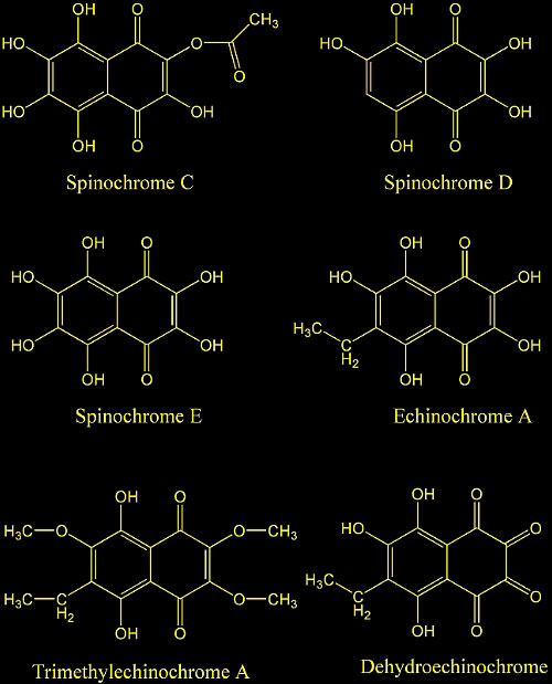
18. Quinoid pigments of echinoderms. VIII. Pigments of sea urchins Diadema setosum and Diadema savignije. Kol'tsova, E. A.; Maksimov, O. B. Tikhookean. Inst. Bioorg. Khim., Vladivostok, USSR. Khimiya Prirodnykh Soedinenii (1981), (1), 115.

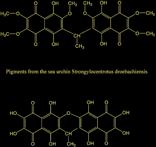
19. Quinoid pigments from echinoderms. V. Pigments from the sea urchin Strongylocentrotus droebachiensis. Kol'tsova, E. A.; Denisenko, V. A.; Maksimov, O. B. Dal'nevost. Nauch. Tsentra, Tikhookean. Inst. Bioorg. Khim., Vladivostok, USSR. Khimiya Prirodnykh Soedinenii (1978), (4), 438-41.

20. Quinoid pigments of echinoderms. III. Minor pigments of the sea urchin Strongylocentrotus nudus. Kol'tsova, E. A.; Chumak, G. N.; Maksimov, O. B. Tikhookean. Inst. Bioorg. Khim., Vladivostok, USSR. Khimiya Prirodnykh Soedinenii (1977), (2), 202-7.

21. Derivatives of naphthoquinones. XIII. Pigments from sea urchins. 8. Kuroda, Chika; Harada, Miye. Sci. Research Inst., Tokyo, Proc. Japan Acad. (1955), 31 305-8.

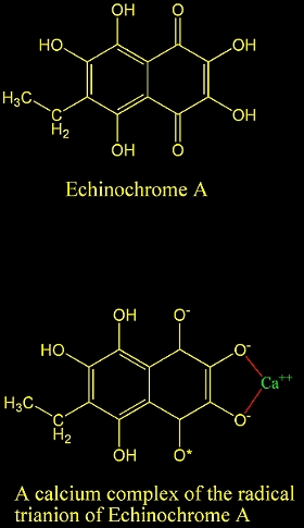
22. How do calcium ions induce free radical oxidation of hydroxy-1,4-naphthoquinone? Ca2+ stabilizes the naphthosemiquinone anion-radical of echinochrome A. Lebedev, Alexander V.; Ivanova, Marina V.; Ruuge, Enno K. Institute of Experimental Cardiology, Department of Biochemistry, Cardiology Research Center, Moscow, Russia. Archives of Biochemistry and Biophysics (2003), 413(2), 191-198.

23. Antifouling agent for fish net. Ashino, Izumi. (Andrews and George Co., Inc., Japan). Jpn. Kokai Tokkyo Koho (1977), 2 pp.

24. Biocidal compositions comprising polyhydroxynaphthoquinones. Sime, Alan Alexander Torrance. (UK). Brit. UK Pat. Appl. (1985), 11 pp.

25. Biocidal compds. for controlling populations of aquatic pest organisms containing quinones, anthraquinones, and naphthalenediones. Cutler, Horace; Cutler, Stephen; Wright, David; Dawson, Rodger. (Garnett, Inc., USA). PCT Int. Appl. (2001), 30 pp.

26. Antimicrobial activity of polyhydroxynaphthoquinones from sea urchins. Stekhova S I; Shentsova E B; Kol'tsova E B; Kulesh N I Antibiotiki i khimioterapiia = Antibiotics and chemoterapy [sic] / Ministerstvo meditsinskoi i mikrobiologicheskoi promyshlennosti SSSR (1988 Nov), 33(11), 831-3.

27. Chemistry of Superoxide Radical in Seawater: CDOM Associated Sink of Superoxide in Coastal Waters. Goldstone, Jared V.; Voelker, Bettina M. Department of Civil and Environmental Engineering, Massachusetts Institute of Technology, Cambridge, MA, USA. Environmental Science and Technology (2000), 34(6), 1043-1048.

28. Chemistry of Superoxide Radical in Seawater: Reactions with Organic Cu Complexes. Voelker, Bettina M.; Sedlak, David L.; Zafiriou, Oliver C. Department of Marine Chemistry and Geochemistry, Woods Hole Oceanographic Institution, Woods Hole, MA, USA. Environmental Science and Technology (2000), 34(6), 1036-1042.

29. Fate of superoxide in coastal sea water. Petasne, Robert G.; Zika, Rod G. Rosenstiel Sch. Mar. Atmos. Sci., Univ. Miami, Miami, FL, USA. Nature (London, United Kingdom) (1987), 325(6104), 516-18.

30. Chemistry of the superoxide radical (O2-) in seawater: Reactions with inorganic copper complexes. Zafiriou, Oliver C.; Voelker, Bettina M.; Sedlak, David L. Woods Hole Oceanographic Institution, Woods Hole, MA, USA. Journal of Physical Chemistry A (1998), 102(28), 5693-5700.

31. Superoxide radical production by sponges Sycon sp. Peskin, Alexander V.; Labas, Yulii A.; Tikhonov, Alexander N. Institute of Developmental Biology, Russian Academy of Sciences, Moscow, Russia. FEBS Letters (1998), 434(1,2), 201-204.

32. Interaction of natural polyhydroxy-1,4-naphthoquinones with superoxide anion-radical. Lebedev, A. V.; Ivanova, M. V.; Krasnovid, N. I. Institute of Experimental Cardiology, Cardiology Research Center, Russian Ministry of Health, Moscow, Russia. Biochemistry (Moscow)(Translation of Biokhimiya (Moscow)) (1999), 64(11), 1273-1278.

33. Mechanisms of inhibition of iron(2+)-induced oxidation of phosphatidylcholine by polyhydroxynaphthoquinones. Lebedev, A. V.; Boguslavskaya, L. V.; Levitskii, D. O.; Maksimov, O. B. Inst. Exp. Cardiol., Cardiol. Res. Cent., Moscow, USSR. Biokhimiya (Moscow) (1988), 53(4), 598-603.

34. Exposure to ultraviolet radiation causes apoptosis in developing sea urchin embryos. Lesser Michael P; Kruse Valerie A; Barry Thomas M Department of Zoology and Center for Marine Biology, University of New Hampshire, Durham, NH 03824, USA. mpl@cisunix.unh.edu Journal of experimental biology (2003 Nov), 206(Pt 22), 4097-103.

35. Synchrotron X-ray studies of the keel of the short-spined sea urchin Lytechinus variegatus: absorption microtomography (microCT) and small beam diffraction mapping. Stock S R; Barss J; Dahl T; Veis A; Almer J D; Carlo F Institute for Bioengineering and Nanoscience in Advanced Medicine, Northwestern University, Chicago, IL 60611, USA. s-stock@northwestern.edu Calcified tissue international (2003 May), 72(5), 555-66.

36. Mineral phase microstructure in teeth of the short spined sea urchin (Lytechinus variegatus) studied with X-ray phase contrast imaging and with absorption microtomography. Stock, S. R.; Dahl, Thomas; Barss, Joseph; Veis, Arthur; Fezzaa, K.; Lee, W. K. School of Materials Sci. & Eng., Georgia Inst. of Technology, Atlanta, GA, USA. Advances in X-Ray Analysis (2002), Volume Date 2001, 45 133-138.

37. Mineral-related proteins of sea urchin teeth: Lytechinus variegatus. Veis Arthur; Barss Joseph; Dahl Thomas; Rahima Mohammed; Stock Stuart Department of Cell and Molecular Biology, Northwestern University Medical School, Chicago, Illinois 60611, USA. aveis@northwestern.edu Microscopy research and technique (2002 Dec 1), 59(5), 342-51.

38. Design strategies of sea urchin teeth: structure, composition and micromechanical relations to function. Wang R Z; Addadi L; Weiner S Department of Structural Biology, Weizmann Institute of Science, Rehorol, Israel Philosophical transactions of the Royal Society of London. Series B, Biological sciences (1997 Apr 29), 352(1352), 469-80.

39. Matrix proteins of the teeth of the sea urchin Lytechinus variegatus. Veis D J; Albinger T M; Clohisy J; Rahima M; Sabsay B; Veis A Journal of experimental zoology (1986 Oct), 240(1), 35-46.

40. Natural carbonate-reinforced composite materials. Part 1. Morphology of sea urchin teeth. Bonfield, W.; Scandola, M. Dep. Mater., Queen Mary Coll., London, UK. Journal of Materials Science (1979), 14(12), 2865-71.

41. X-ray absorption microtomography (microCT) and small beam diffraction mapping of sea urchin teeth. Stock S R; Barss J; Dahl T; Veis A; Almer J D Institute for Bioengineering and Nanoscience in Advanced Medicine, Northwestern University, Chicago, IL 60611-3008, USA. s-stock@northwestern.edu Journal of structural biology (2002 Jul), 139(1), 1-12.

42. Recent Studies on the Pathological Effects of Purified Sea Urchin Toxins. Nakagawa, H.; Tanigawa, T.; Tomita, K.; Tomihara, Y.; Araki, Y.; Tachikawa, E. Department of Life Sciences, University of Tokushima, Tokushima, Japan. Journal of Toxicology, Toxin Reviews (2003), 22(4), 633-649.

43. The inhibitory effect of the toxic fraction from sea urchin (Toxopneustes pileolus) venom on 45Ca2+ uptake in crude synaptosome fraction from chick brain. Zhang, Yu-An; Wada, Tetsuyuki; Iwasaki, Yasunori; Nakagawa, Hideyuki; Ichida, Seiji. Department of Biological Chemistry, Faculty of Pharmaceutical Sciences, Kinki University, Higashi-Osaka, Japan. Biological & Pharmaceutical Bulletin (1999), 22(12), 1279-1283.

44. Mode of attack of sea urchin toxin on natural and synthetic substrates. II. Physical properties, substrate specificity, and reaction kinetics of purified fractions. Feigen, George A.; Hadji, Lahlou; Pfeffer, Roger A.; Markus, Gabor. Dep. Physiol., Stanford Univ., Stanford, CA, USA. Physiological Chemistry and Physics (1970), 2(5), 427-44.

45. Fishing for bioactive substances from scorpionfish and some sea urchins. Satoh, F.; Nakagawa, H.; Yamada, H.; Nagasaka, K.; Nagasaka, T.; Araki, Y.; Tomihara, Y.; Nozaki, M.; Sakuraba, H.; Ohshima, T.; Hatakeyama, T.; Aoyagi, H. Department of Life Sciences, University of Tokushima, Tokushima, Japan. Journal of Natural Toxins (2002), 11(4), 297-304.

46. Purification and characterization of contractin A from the pedicellarial venom of sea urchin, Toxopneustes pileolus. Nakagawa, Hideyuki; Tu, Anthony T.; Kimura, Akira. Dep. Biochem., Colorado State Univ., Fort Collins, CO, USA. Archives of Biochemistry and Biophysics (1991), 284(2), 279-84.

47. Effect of Contractin A, a glycoprotein from the pedicellarial venom of the sea urchin Toxopneustes pileolus, on isolated vascular smooth muscles. Nakagawa, Hideyuki; Kitagawa, Hisato; Kondo, Noriaki; Kondo, Jun. Fac. Integrated Arts Sci., Univ. Tokushima, Tokushima, Japan. Journal of Natural Toxins (1992), 1(2), 31-7. CODEN: JNTOER ISSN: 1058-8108.

48. Preliminary studies on venom proteins in the pedicellariae of the toxopneustid sea urchins, Toxopneustes pileolus and Tripneustes gratilla. Nakagawa, Hideyuki; Tomihara, Yasuhiro; Araki, Yasutetsu; Hayashi, Hiromi. Dep. Life Sci., Univ. Tokushima, Tokushima, Japan. Journal of Natural Toxins (1994), 3(1), 25-34.


Reefkeeping Magazine™ Reef Central, LLC-Copyright © 2008

Sea Urchins: A Chemical Perspective by Randy Holmes-Farley - Reefkeeping.com